Effect of Space Flight on Bone Quality using High Resolution Imaging
Studying astronauts’ bones after months in space provides an understanding of bone disease here on Earth, equivalent to years of aging
After the age of 50, we typically lose 1 per cent of our bone mass every year, a process that can lead to osteoporosis, a bone loss disorder that affects about ten per cent of Canadians aged 40 and over. In space, with weightlessness and reduced loading, astronauts can lose up to 1.5 per cent of their bone mass every month. Although some of it returns when they’re back on Earth, its structure is different.
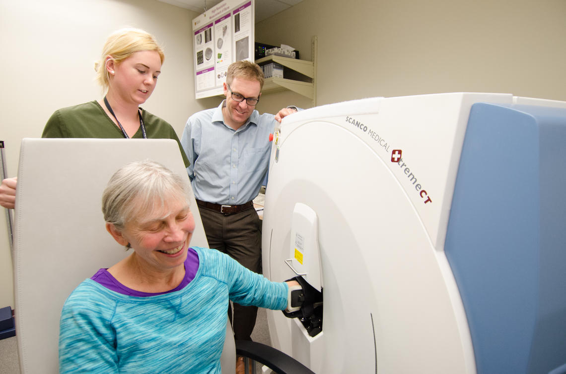
BME researchers are working with astronauts to understand that cycle. They are measuring changes to the interior structure of bones in astronauts using high resolution peripheral quantitative computed tomography (HR-pQCT) and novel image analysis techniques. The astronauts are assessed after they have spent six months on the International Space Station and their recovery is monitored for a year after they have returned. This information will further the understanding of the mechanisms of loss and repair of bone microarchitecture. It will have implications for astronauts in long-term space fight and help us understand how to prevent osteoporosis here on Earth. The study will help identify people at risk of bone loss and help come up with ways to predict and prevent fractures caused by low bone density.

Participant getting their forearm (radius bone) scanned using HR-pQCT at the McCaig Institute
In the News

3D high resolution peripheral quantitative computed tomography (HR-pQCT) scanner
Paul Hulme
Stephanie Kwong
Partners
Funded by the Canadian Space Agency
NASA – Scott Smith, Jean Sibonga
European Space Agency - Anna-Maria Liphardt, Martina Heer


